×


Accessories
Acquisition Systems
Catheters
CT Scanners
DR Detectors
Generators
Interventional X-Ray
Mammography
Medical Tables
Mobile X-Ray
Monitors
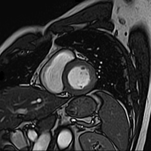
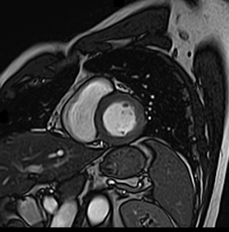
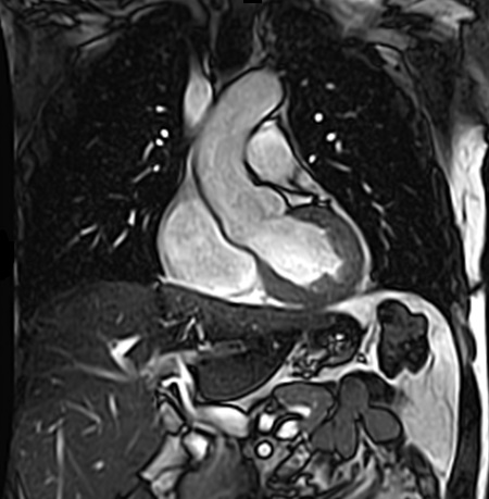
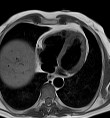
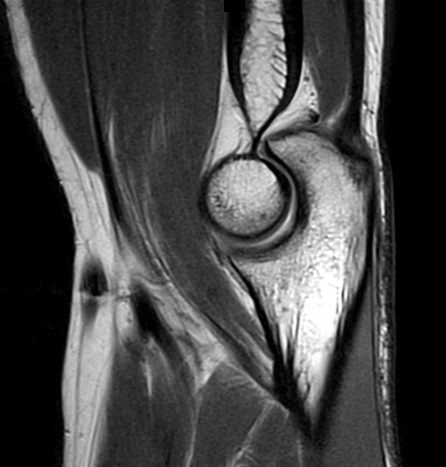
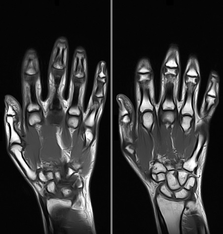
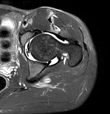
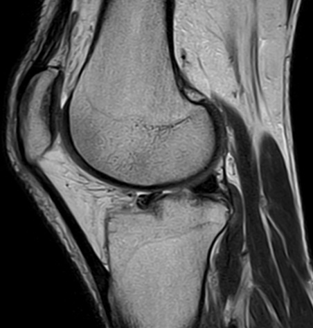
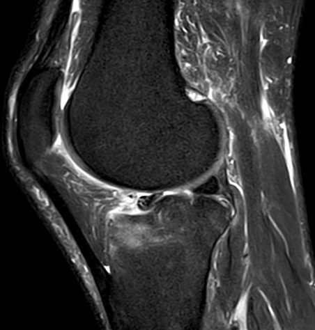
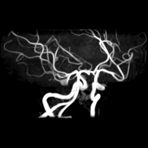
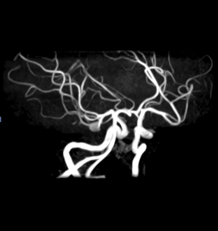
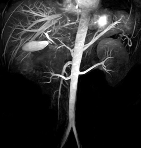
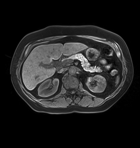
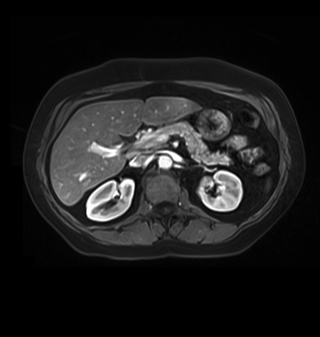
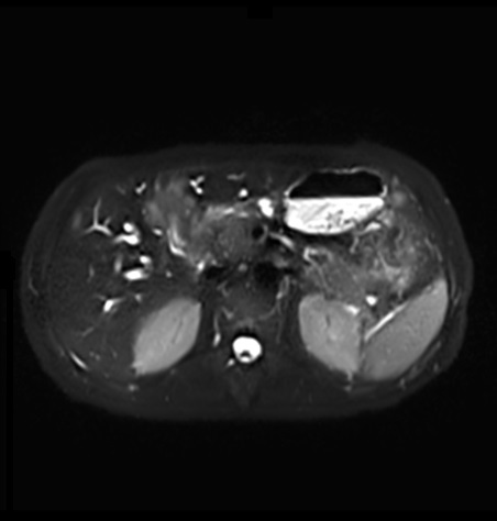
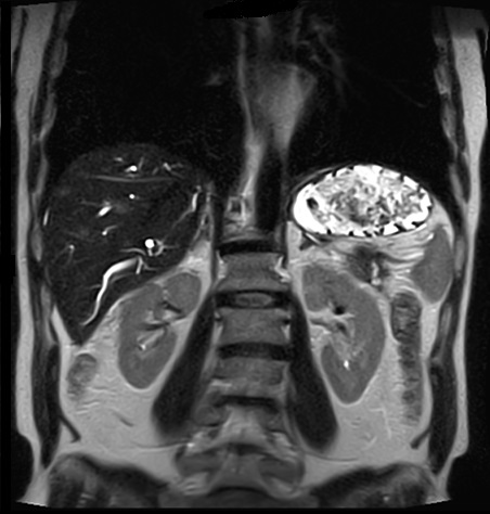
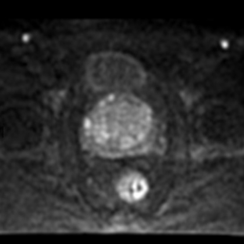
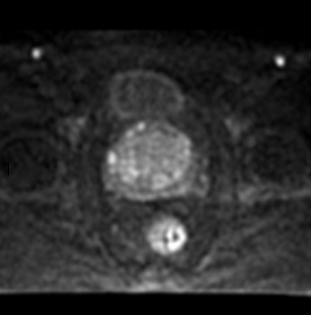
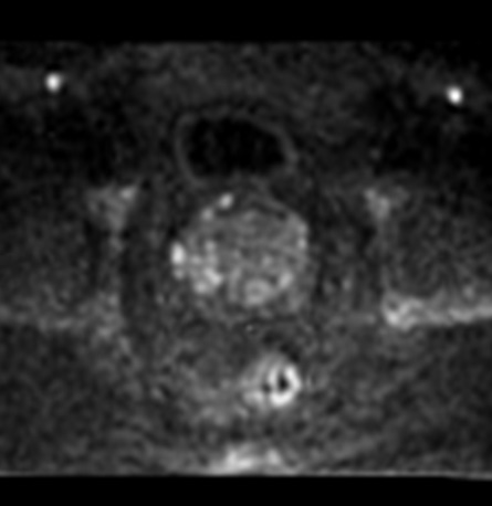
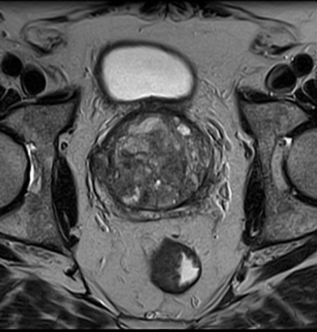
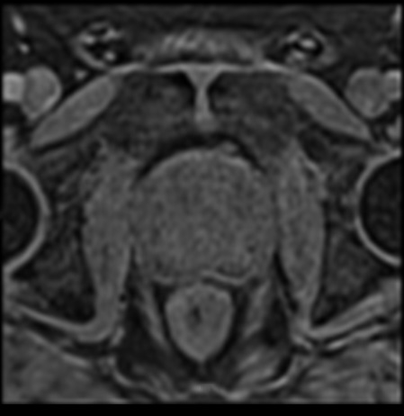
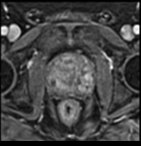
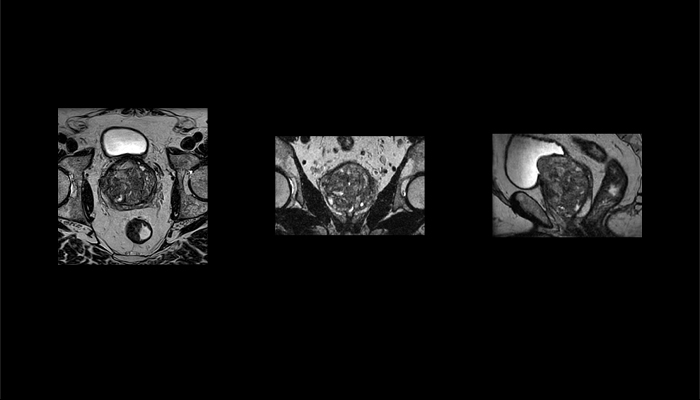
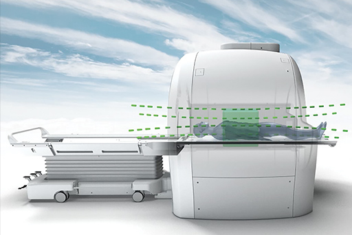
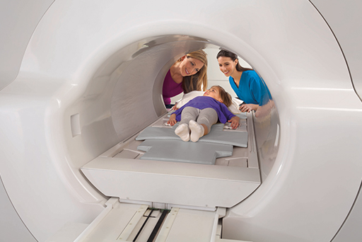
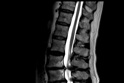
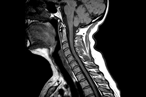
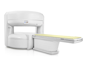
MRI
Office Based Laboratory
Overhead Tube Cranes & Tube Stands
Radiography / Fluoroscopy
Software / Informatics
Straight, C, & U Arms
Upgrades
Wallstands
Shimadzu
Canon
Konica Minolta
UMG Del Medical
Medlink
Cuattro
Eizo
Wide
Fuji
Avreo GE Healthcare - EdisonTM True PACS
MedCurrent
Gen 7 Healthcare
RC Imaging
Techno-Aide
Bar-Ray
SealCath
Accessories
Installations


 ©
- CMS Imaging, Inc. All
Rights
Reserved
©
- CMS Imaging, Inc. All
Rights
Reserved
